Eye anatomy, artwork Wall Art
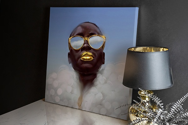
12″ × 16″ Stretched Canvas Print
About the Artwork
Eye anatomy, artwork - Item # 1154218
Eye anatomy. Artwork of a section through part of a healthy human eyeball. The cornea (the transparent outer part of the front of the eye) runs from centre to bottom right. Immediately behind this is the aqueous humour, a liquid that supports the structures. This liquid flows through the eye, as shown by the red arrows. The dense mesh at lower centre is part of the iris, the ring-shaped muscle that surrounds the pupil (not seen). To the left of the iris is the ciliary body (pink), from which the iris is derived. At bottom left, thin zonule filaments are seen. These hold the lens (not seen) of the eye in place behind the pupil.
Product Specifications
- Expertly Handcrafted
- 1.25" Solid Wood Stretcher Bars
- Artist-Grade Canvas
- Fade-Resistant Archival Inks
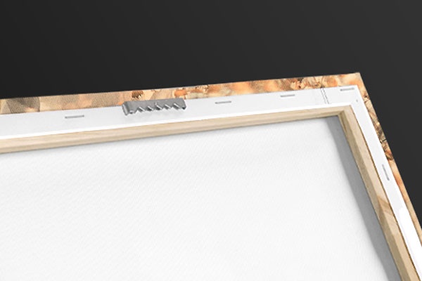
- Hanging Hardware Pre-Installed
- Width: 12″
- Height: 16″
Item # 1154218
Product Features
Elevate any room with our handcrafted stretched canvas gallery wraps. Printed with archival inks and wrapped around a 1.25” inch solid wood stretcher bar, our giclée big canvas art prints are a timeless option for any decor style or space.

Our giclée canvas art prints are produced with high quality, UV-resistant, environmentally-friendly, latex inks and artist grade, polycotton canvas. We pride ourselves on color accuracy and image clarity to ensure your new canvas wall art lasts for years to come.

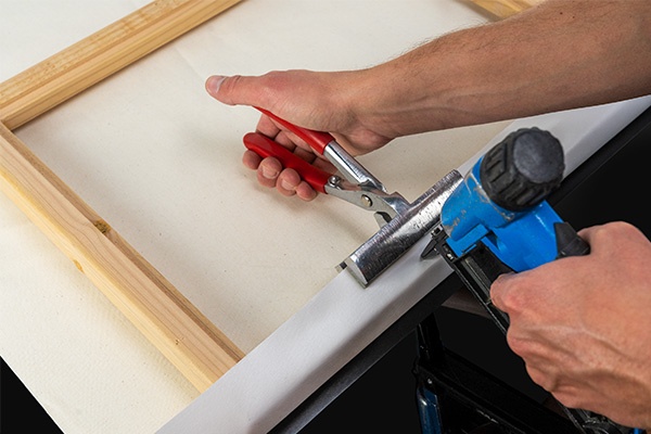
Assembled in the USA, each of our 1.25” inch gallery wrapped canvas art prints is stretched and stapled by our highly skilled craftspeople. Each canvas print is carefully handcrafted to ensure taut canvas wraps and clean corners for outstanding quality and durability.

Our handcrafted stretched canvas prints include sawtooth hangers for an easy and secure installation.
Product Reviews
Frequent Questions
Recently Viewed
Clear Recently Viewed?
Are you sure you would like to clear your recently viewed items?
Pricing policy: The full list price is a price at which we have offered the product for sale; however, we may not have sold the item at that price.

