Heel skin tissue, light micrograph Wall Art
12″ × 18″ Stretched Canvas Print
About the Artwork
Heel skin tissue, light micrograph - Item # 1145921
Heel skin tissue. Polarised light micrograph of a transverse section through skin from the heel of a human foot. The sole of the foot has to withstand the weight of the body and friction when the foot pushes against the substratum. To cope with this, the outer epidermis forms deep layers of dead keratinised cells (red and blue) which are replaced from the meristematic Malpighian layer as they get worn away. The dermis (yellow and green) consists of collagenous elastic fibres. Magnification: x103 when printed at 10 centimetres high.
Product Specifications
- Expertly Handcrafted
- 1.25" Solid Wood Stretcher Bars
- Artist-Grade Canvas
- Fade-Resistant Archival Inks
- Hanging Hardware Pre-Installed
- Width: 12″
- Height: 18″
Item # 1145921
Product Features
Elevate any room with our handcrafted stretched canvas gallery wraps. Printed with archival inks and wrapped around a 1.25” inch solid wood stretcher bar, our giclée big canvas art prints are a timeless option for any decor style or space.

Our giclée canvas art prints are produced with high quality, UV-resistant, environmentally-friendly, latex inks and artist grade, polycotton canvas. We pride ourselves on color accuracy and image clarity to ensure your new canvas wall art lasts for years to come.

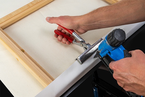
Assembled in the USA, each of our 1.25” inch gallery wrapped canvas art prints is stretched and stapled by our highly skilled craftspeople. Each canvas print is carefully handcrafted to ensure taut canvas wraps and clean corners for outstanding quality and durability.

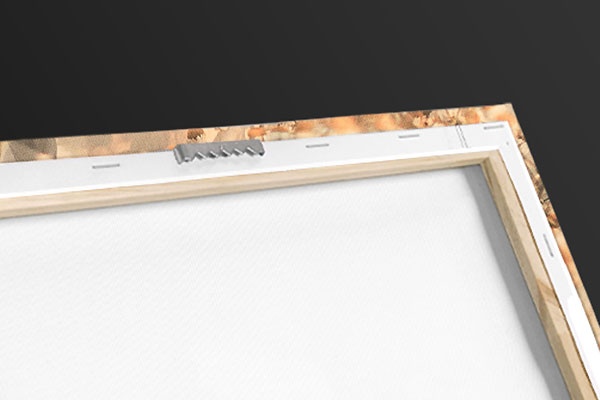
Our handcrafted stretched canvas prints include sawtooth hangers for an easy and secure installation.
Product Reviews
Frequent Questions
Recently Viewed
Clear Recently Viewed?
Are you sure you would like to clear your recently viewed items?
Pricing policy: The full list price is a price at which we have offered the product for sale; however, we may not have sold the item at that price.

